
Sometimes science can be a messy endeavor—not to mention "disgusting and smelly." That's how British researchers described their experiments monitoring dead sea bass carcasses as they rotted over the course of 70 days. In the process, they gained some fascinating insights into how (and why) the soft tissues of internal organs can be selectively preserved in the fossil record, according to a new paper published in the journal Palaeontology.
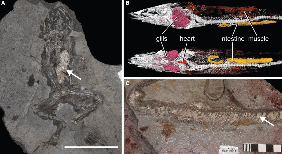
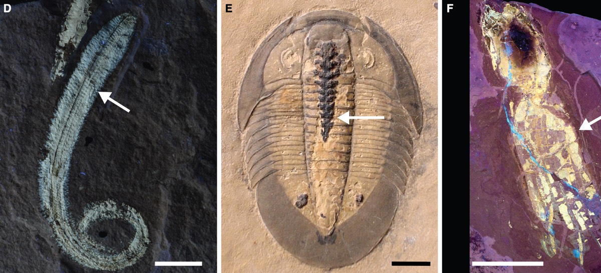
Most fossils are bone, shells, teeth, and other forms of "hard" tissue, but occasionally rare fossils are discovered that preserve soft tissues like skin, muscles, organs, or even the occasional eyeball. This can tell scientists much about aspects of the biology, ecology, and evolution of such ancient organisms that skeletons alone can't convey. For instance, earlier this year, researchers created a highly detailed 3D model of a 365-million-year-old ammonite fossil from the Jurassic period by combining advanced imaging techniques, revealing internal muscles that had never been previously observed.
"One of the best ways that soft tissue can turn into rock is when they are replaced by a mineral called calcium phosphate (sometimes called apatite)," said co-author Thomas Clements of the University of Birmingham. "Scientists have been studying calcium phosphate for decades trying to understand how this process happens—but one question we just don’t understand is why some internal organs seem more likely to be preserved than others."
Specifically, muscles, stomachs, and intestines tend to "phosphatize" much more frequently than other organs, like kidneys and gonads. There are two common hypotheses to explain this. The first is that different organs decay at different rates, and the pH of certain organs will fall below the critical threshold of 6.4. As these organs decay, they create a distinct pH microenvironment that increases the likelihood of those organs being fossilized. Different minerals may form in different areas within the same carcass.
The second hypothesis is that tissue biochemistry plays a major role. Specifically, a pervasive pH environment forms within the body cavity and persists until the carcass breaks down.
According to Clement et al., no prior research has focused on documenting the pH gradients associated with the decay of specific anatomical features as a carcass rots in real time; past experiments have focused on recording pH fluctuations outside the carcass. So the team decided to rectify that gap and conduct experiments on decaying fish, documenting how the pH gradient changed over the course of two-and-a-half months.
First, they purchased several adult European sea bass from a local fishmonger as soon after death as possible (no more than 24-36 hours). The fish were kept on ice to slow down decay but were not frozen to avoid any cellular damage. Next, they inserted pH probes into various locations on each of the six sea bass carcasses to target specific organs: stomach, liver, intestines, and epaxial muscle. A fifth probe was used to monitor the pH of the surrounding environment between 1 and 2 mm away from the carcass.
reader comments
71